16 million-year-old amber specimen reveals unknown animal behaviours
18 Oct 2012
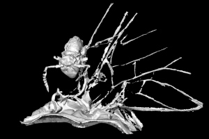
Stunning images, including video footage, from a CT scan of amber have revealed the first evidence of any creature using an adult mayfly for transport. Researchers at the University of Manchester say this 16 million-year-old hitchhiker most likely demonstrates activity that is taking place today but has never previously been recorded.

Entombed in amber the tiny springtail can be seen resting in a v-shaped depression at the base of one of the mayfly's wings. It appears to have secured itself for transport using its prehensile antennae.
Dr David Penney and colleagues from the Faculty of Life Sciences and the School of Materials used a high resolution CT scanner to take over 3,000 X-rays from different angles. The scientists then created slices, showing the fossil in cross sections. From these slices 3D digital images of the springtail were made so an accurate analysis of its behaviour could be conducted.
Dr Penney says: “The images are really impressive. This pioneering approach to studying fossils has allowed us an insight into the behaviour of one of the world’s most prevalent organisms.”
Springtails are minute creatures (usually only 1-2mm long) related to true insects. They’re found around the world in great numbers, including here in the UK. Gardeners will recognise them as the tiny insects that hop around when soil is disturbed. They readily colonize newly-formed islands but very little is known about how they manage to migrate. One of the reasons is that they are incredibly nervous creatures and have an astonishing ability to leap away from danger using a springing organ (the furca) on the underside of the abdomen, which makes observing them in life very difficult.
Interestingly, when the 3D image of the springtail in amber is magnified it's possible to see that the springtail is very slightly detached (by just 50 micrometres) from the mayfly. This suggests it was attempting to spring away as the amber set around it.
Only one previous case of phoresy (the transportation of one organism by another) has been recorded for springtails. This was found in a piece of Baltic amber where five springtails were hooked in a row on the leg of a harvestman arachnid.
It was this discovery in 2010 which prompted Dr Penney to take a closer look at his own specimen. “I had initially thought the creature on the mayfly may have been a tiny nymphal pseudoscorpion, as they are known to use other creatures for transport, and this behaviour is not uncommon to see in amber. I was interested in the fact that this was the first time a creature had been found on an adult mayfly but I didn't truly appreciate the significance of my find until I used the CT scanner and was able to identify the animal as a springtail.”
Phoresy in adult mayflies has never before been recorded. They live for just a short period of time from one hour to a few days depending on the species. The primary function of the adult stage is reproduction and they are unable to feed. This makes it very difficult to study mayflies in their natural habitat and record instances of phoresy.
The amber specimen encasing the mayfly and the springtail provides an accurate snapshot of behaviour that scientists wouldn't otherwise be able to record, highlighting one important application of the fossil record for understanding the present. The near perfect condition of the mayfly demonstrates that it died instantaneously and wasn't moved far from where it rested when the resin ran over it. Equally the position of the springtail resting on the back of the mayfly and the fact that it is in contact with the creature means the pair were unlikely to have been brought together by the resin as it moved down the tree.
The details of Dr Penney’s research has been published in the journal, PLOS ONE. More analysis of amber using CT scans is continuing.
Dr Penney says: “The CT scan allows us to build up a 3D image that catches minute details of the animal. We can rotate the image to see parts of the creature that are obscured when looking from the outside in. In effect, we are able to digitally dissect the fossil without causing any damage to it whatsoever. This technology has revolutionised how we study fossils and the findings are incredibly exciting.”
