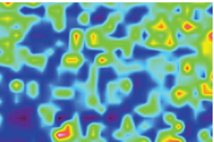
First image of protein residue in 50 million year old reptile skin
23 Mar 2011
The organic compounds surviving in fifty million year old fossilized reptile skin can be seen for the first time today, thanks to a stunning infra-red image produced by University of Manchester palaeontologists and geochemists.

Published in the journal Royal Society Proceedings B, the brightly-coloured image shows the presence of amides – the organic compounds, or building blocks of life – in the ancient skin of a reptile, found in the 50 million year-old rocks of the Green River Formation in Utah, USA.
This image had never been seen by the human eye, until a team led by Dr Roy Wogelius and Dr Phil Manning used state-of-the-art infra-red technology at The University of Manchester to reveal and map the fossilized soft tissue of a beautifully-preserved reptile.
These infra-red maps are backed up by the first ever element-specific maps of organic material in fossil skin generated using X-rays at the Stanford synchrotron in the USA, also by the Manchester researchers.
Chemical details are clear enough that the scientists, from the School of Earth, Atmospheric and Environmental Sciences, are even able to propose how this exceptional preservation occurs.
When the original compounds in the skin begin to break down they can form chemical bonds with trace metals, and under exceptional conditions these trace metals act like a ‘bridge’ to minerals in the sediments. This protects the skin material from being washed away or decomposing further.
Geochemist Roy Wogelius: “The mapped distributions of organic compounds and trace metals in 50 million year old skin look so much like maps we’ve made of modern lizard skin as a check on our work, it is sometimes hard to tell which is the fossil and which is fresh.”
“These new infra-red and X-ray methods reveal intricate chemical patterns that have been overlooked by traditional methods for decades.”
The new images are compelling, and represent the next step in the academics’ research programme to use modern analytical chemistry and 21st century techniques to understand how such remarkable preservation occurs, and ultimately to discover the chemistry of ancient life.
These new results imply that trace metal inventories and patterns in ancient reptile skin, even after fossilisation, can indeed be compared to modern reptiles.
The infra-red light causes vibrations in the fossilized skin, and a map of where these vibrations occur can be obtained from a fossil by using a trick: a tiny crystal (like an old phonograph record stylus) which moves from point-to-point in a programmable grid across the surface.
At each point where the tiny crystal touches the fossil, an infra-red beam that shines through the crystal reflects off of the crystal base, but a small amount of the beam probes beyond the interface- and if organic compounds are present, they absorb portions of the beam and change the reflected signal.
This allows the team to non-destructively map large fossils which do not themselves transmit or reflect the beam – a revolutionary process for paleontologists.
Nick Edwards, first author on the publication, said: “The ability to chemically analyse rare and precious fossils such as these without the need to remove material and destroy them is an important and long overdue addition to field of palaeontology.
“Hopefully this will provide future opportunities to unlock the information stored in other similarly preserved specimens.”
Dr Manning said: “Here physics, palaeontology and chemistry have collided to yield incredible insight to the building blocks of fossilized soft tissue.
“The results of this study have wider implications, such as understanding what happens to buried wastes over long periods of time. The fossil record provides us with a long-running experiment, from which we can learn in order to help resolve current problems.”
